What Happens When an ACL Tear Comes With a Fracture

An ACL tear with fracture is more common than most people realize — and missing the fracture part can seriously set back your recovery.
Here's a quick breakdown of what you need to know:
| Key Fact | Detail |
|---|---|
| How common are fractures with ACL tears? | 60–72% of acute ACL tears show fractures on MRI |
| Most recognizable fracture type | Segond fracture — seen on X-ray, linked to ACL tear in 75–100% of cases |
| Most common missed fracture type | Subchondral (bone surface) fractures — invisible on X-ray, visible on MRI |
| Why it matters | Associated fractures increase the risk of meniscal tears and joint damage |
| Who is most affected? | Athletes, active adults, and children aged 8–14 (avulsion fractures) |
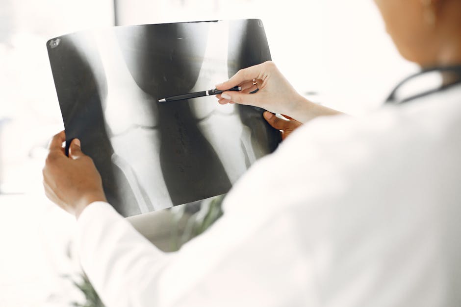
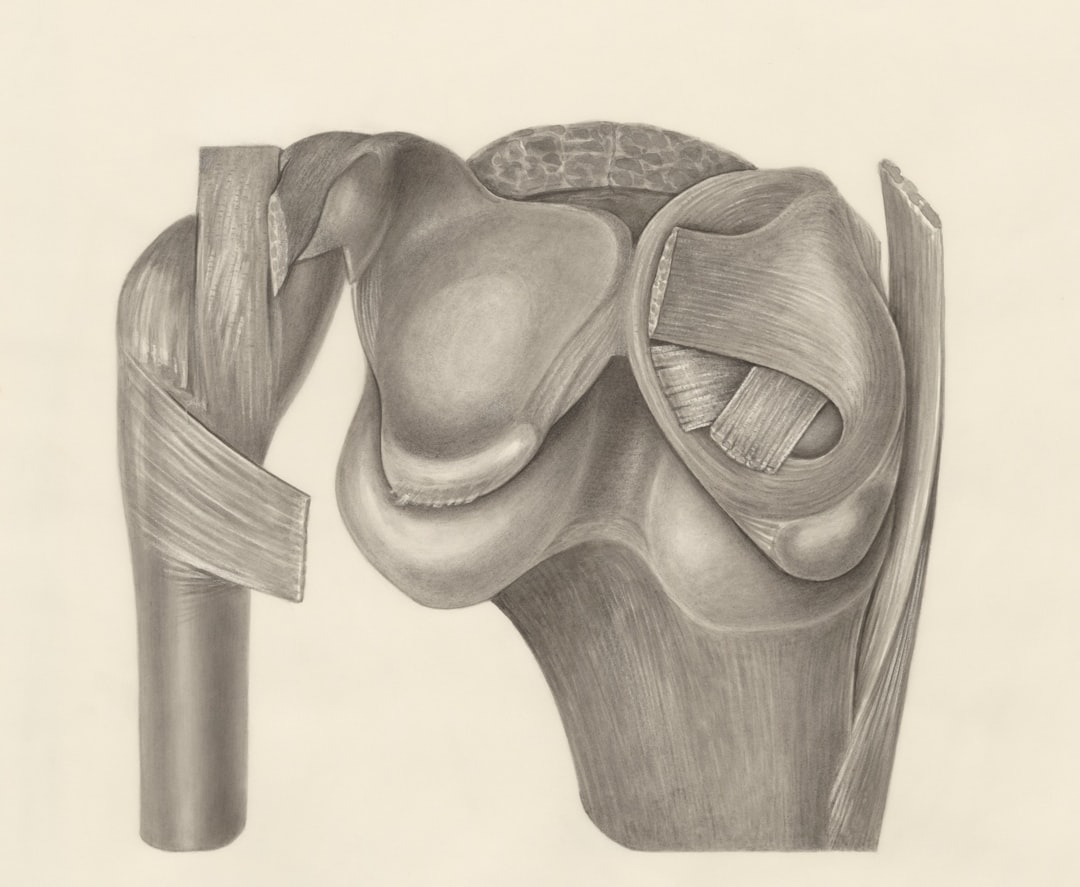
The ACL (anterior cruciate ligament) connects your thighbone (femur) to your shinbone (tibia). It keeps your knee stable during cutting, pivoting, and landing movements. Around 200,000 Americans tear this ligament every year — and in a large portion of those cases, at least one bone injury comes along with it.
The problem? Standard X-rays often miss the fractures entirely. That means patients walk away with an incomplete diagnosis — and sometimes, an incomplete recovery.
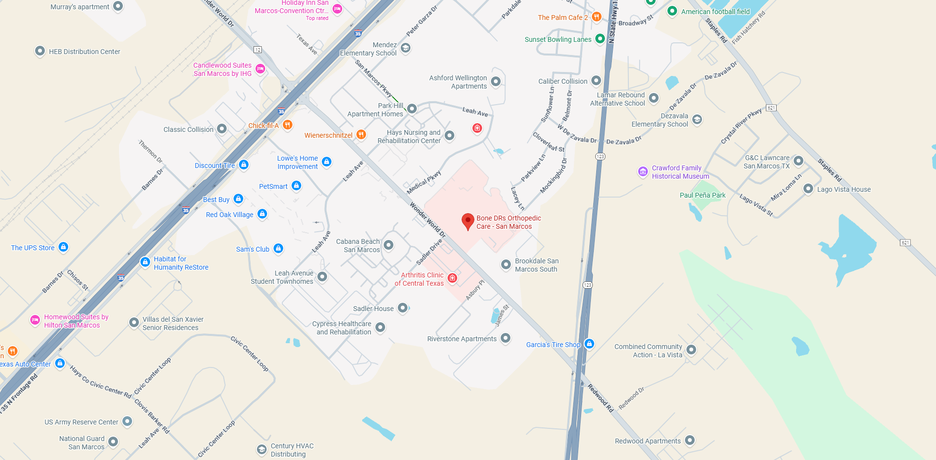
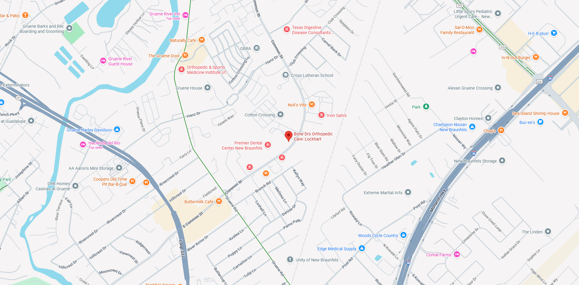
I'm Christopher Jimenez, MD, a board-certified orthopedic surgeon with extensive experience treating lower extremity trauma, including complex cases of ACL tear with fracture. Managing these combined injuries — from initial diagnosis to surgical reconstruction — is a core part of what I do at Bone Drs Orthopedic Care in Central Texas.

Common Types of ACL Tear With Fracture
When we talk about an ACL tear with fracture, we aren't usually talking about a snapped leg bone that requires a full cast. Instead, these are often "avulsion" fractures—where the ligament pulls a piece of bone away—or "impaction" fractures, where the bones slam into each other during the injury.
Research indicates that fractures associated with ACL injury need to be taken seriously because they change the environment of the knee joint. At BONE DRs, our fractures & trauma services focus on identifying these subtle bone injuries that can complicate a standard ligament repair.
The most common types include:
- Segond Fracture: A tiny chip of bone off the outer edge of the tibia.
- Tibial Eminence Avulsion: The "root" of the ACL pulls a chunk of bone out of the shinbone.
- Subchondral Fractures: Cracks just beneath the joint surface, often appearing as severe "bone bruising."
- Lateral Femoral Condyle Impaction: A dent in the thighbone caused by the tibia hitting it during a pivot-shift movement.
The Segond Fracture: A Red Flag for ACL Injury
The Segond fracture is perhaps the most famous "sidekick" to an ACL injury. Named after Paul Segond, who described it in 1879, it is a small avulsion fracture of the lateral tibial plateau.
Why do we care so much about a tiny sliver of bone? Because it is considered pathognomonic for an ACL rupture. This means if we see a Segond fracture on your X-ray, there is a 75% to 100% chance you also have a torn ACL. It occurs when the tension on the lateral side of the knee—specifically involving the anterolateral ligament (ALL)—becomes so great during a twisting motion that it rips a piece of bone right off the tibia.

Identifying this "red flag" early helps us provide a more accurate diagnosis of knee ligament injuries before you even step into the MRI suite.
Tibial Eminence Avulsions in Pediatric Patients
In our younger patients in Austin and San Marcos, we see a different pattern. Between the ages of 8 and 14, a child’s ligaments are often stronger than their developing bones. Instead of the ligament fibers snapping in the middle (a mid-substance tear), the ACL stays intact but pulls its bony attachment point—the tibial eminence—away from the rest of the shinbone.
We use the Meyers-McKeever classification to grade these:
- Type I: The bone fragment is barely moved (non-displaced).
- Type II: The front of the fragment is lifted like a "beak."
- Type III: The entire bone fragment is floating free.
Because these involve the growth plates and the structural integrity of the tibia, they require specialized pediatric orthopedic attention to ensure the knee grows correctly.
Subchondral and Impaction Fractures in an ACL Tear With Fracture
While Segond fractures get the headlines, subchondral fractures are much more common. Research shows that 60% to 72% of acute ACL tears involve these fractures.
Unlike a clean break, these are often "impaction" injuries. Imagine two cars colliding; even if the frames don't snap, the bumpers get dented. In the knee, the lateral femoral condyle (thighbone) and the posterolateral tibial plateau (shinbone) slam into each other during the "pivot-shift" moment of the injury. This creates a deep "bone bruise" or a subchondral fracture.
The Biomechanics of Combined Knee Injuries
How does an ACL tear with fracture actually happen? It’s usually a perfect storm of forces. Most ACL injuries (over 70%) are non-contact, occurring during a sudden deceleration or a "pivot-shift."
When you plant your foot and twist, several things happen at once:
- Valgus Stress: The knee collapses inward.
- Internal Rotation: The tibia twists relative to the femur.
- Anterior Translation: The tibia slides forward because the ACL is no longer holding it back.
This violent shift is what causes the bones to knock together (causing impaction fractures) or pulls the ligaments so tight they snap bone off (causing avulsion fractures). If you're experiencing significant knee pain after a "pop," these biomechanical forces are likely the culprit.
Impact of ACL Tear With Fracture on Meniscal Health
A bone fracture isn't the only thing that hitches a ride with an ACL tear. The meniscus—the shock-absorbing cartilage in your knee—is frequently caught in the crossfire.
In cases where a Segond fracture is present, the incidence of lateral meniscus tears jumps significantly. One study found that isolated lateral meniscus tears occurred in 50% of ACL ruptures with a Segond fracture, compared to only 20.8% in those without.
We also look closely for:
- Posterior Root Tears: Where the meniscus detaches from the bone.
- Medial Ramp Lesions: Hidden tears at the back of the medial meniscus that are present in nearly 40% of ACL cases but are often missed during standard surgery.
Detailed MRI analysis is essential to catch these, as leaving a ramp lesion or root tear untreated can lead to early-onset osteoarthritis and even cause an ACL graft to fail.
The New Injury Tetrad: High-Velocity Trauma
In high-velocity accidents—like a motorcycle fall—we sometimes see a devastating combination known as the "New Injury Tetrad." This involves four major injuries at once:
- ACL Rupture
- MCL (Medial Collateral Ligament) Tear
- Lateral Meniscus Posterior Root Tear
- Posterolateral Tibia Osteochondral Fracture
This is a complex scenario that requires a "fracture-first" mindset. We must stabilize the bone fracture before we can successfully reconstruct the ligaments.
Diagnostic Imaging: Beyond the Standard X-Ray
If you go to an urgent care in Bastrop or Lockhart after a knee injury, they will likely start with an X-ray. While X-rays are great for seeing a Segond fracture or a displaced tibial eminence, they are notoriously bad at seeing the most common fractures associated with ACL tears.
This is where MRI becomes the "gold standard." MRI allows us to see:
- Bone Marrow Edema: The "bruising" that indicates where the bones collided.
- T1-Weighted Dark Lines: These represent the actual fracture lines in the subchondral bone.
- Soft Tissue Detail: Essential for planning a knee arthroscopy.
Identifying an ACL Tear With Fracture on MRI
On an MRI, a fracture looks different than a simple bruise. A "bone bruise" is an ill-defined area of swelling (edema). A fracture, however, shows up as a distinct, dark, low-signal intensity line.
Detecting these "occult" (hidden) fractures is vital. If we find an impaction fracture on the lateral femoral condyle, it tells us exactly how the injury happened and warns us that the surrounding cartilage may be damaged as well.
Treatment Strategies and Surgical Considerations
When dealing with an ACL tear with fracture, the treatment plan gets a bit more "custom-made." We don't just fix the ligament; we have to address the "neighborhood" it lives in.
For most active patients, ACL reconstruction is the primary goal. However, if there is a significant fracture, we may use:
- Arthroscopic Staple Fixation: Used for tibial eminence fractures to "tack" the bone fragment back into place.
- Headless Compression Screws: These tiny screws sink below the surface of the bone/cartilage to hold fragments together without irritating the joint.
- Simultaneous Repair: We often perform meniscus surgery and knee ligament reconstruction in the same session to ensure total stability.
Managing a Pediatric ACL Tear With Fracture
For kids with a tibial eminence avulsion, we have a unique opportunity. Because the ligament itself is often still healthy, we can sometimes avoid a full reconstruction.
If the fracture is not displaced (Type I), we might use non-operative treatment, such as immobilizing the knee in extension for several weeks. If it is displaced, we perform a minimally invasive surgery to pull the bone fragment back down and secure it. This preserves the child's natural ACL and protects their growth plates, which is a key consideration in our patellar tendon repair and pediatric protocols.
Frequently Asked Questions about ACL Fractures
Can an ACL tear cause a bone fracture?
Yes, absolutely. Because the ACL is anchored directly into the bone, the force required to tear the ligament can sometimes be enough to pull a piece of bone away (an avulsion fracture) or cause the bones of the knee to slam together (an impaction fracture).
How does a Segond fracture affect ACL surgery?
A Segond fracture serves as a warning that the injury was high-energy and that the lateral side of the knee is unstable. While the fracture itself often heals on its own once the knee is stabilized, its presence may lead your surgeon to check more closely for lateral meniscus tears or to consider reinforcing the lateral side of the knee during your ACL reconstruction.
What is the recovery timeline for an ACL tear with fracture?
Adding a fracture to an ACL tear can sometimes slightly delay the "weight-bearing" phase of recovery. While a standard ACL patient might be walking within a week or two, a patient with a tibial plateau fracture might need to be on crutches for 4 to 6 weeks to allow the bone to knit back together. However, the total return-to-sport timeline usually remains around 6 to 12 months.
Conclusion
An ACL tear with fracture is a complex injury that requires a keen eye and a comprehensive treatment plan. Whether it’s a Segond fracture acting as a diagnostic "red flag" or a subchondral fracture that explains your deep joint pain, these bone injuries are a critical part of the story.
At BONE DRs, we believe in treating the whole knee, not just the ligament. By using advanced imaging and personalized surgical techniques, we help patients in Austin, San Marcos, Bastrop, and North Austin get back to the activities they love with a knee that is stable from the bone up.
If you’ve suffered a knee injury and want to ensure no part of your diagnosis is missed, schedule a consultation for fractures and trauma with our team today. We’re here to help you navigate the road to recovery—fractures and all.
If you have any concerns about your joints, contact us and we can help determine a treatment that works for you.